The Magnetic Power of the Future: The Iseult CEA 11.7 T MRI
Published on: December 16, 2024
What is the Iseult CEA 11.7 T MRI?
The Iseult CEA 11.7 Tesla (T) MRI machine marks a significant leap forward in magnetic resonance imaging, offering unmatched magnetic field strength. It was built through a collaborative effort from the French Alternative Energies and Atomic Energy Commission (CEA), together with Alstrom Magnets and Superconductors and Siemens Healthineers, all under the umbrella and framework of the French-German consortium Iseult. This MRI machine operates at a strength of 11.7 T, making it the most powerful MRI machine for human imaging produced thus far. This feat marks the culmination of nearly 20 years of research and development under the Iseult project, with the goal of building a “human brain explorer” that investigates the brain at an unprecedented scale.
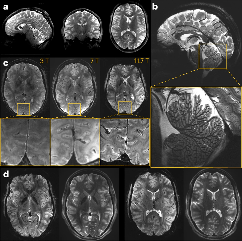
The first in vivo human brain images using this MRI were unveiled on April 2, 2024, and they are game-changing. Compared to standard 3T and even 7T MRIs, the 11.7 T images boast a vastly higher signal-to-noise ratio (SNR), enabling superior spatial and temporal resolution. Within just 5 minutes of scanning, this machine achieved an incredible resolution of 0.19 mm in-plane and 1 mm slice thickness (0.19 x 0.19 x 1 mm³). To put this into perspective, a typical 3T MRI would need around 15 times longer to reach a similar resolution—an impractical timeframe for clinical use.
The specs of the 11.7 T MRI include a 5 m long and 5 m tall cylinder, a magnet that weighs a whopping 132 tons in total, 182 km of superconducting wires, and 7,500 L of superfluid liquid helium to cool the magnet at -271.35C. Achieving the specs underlying this machine required new methodology specifically related to the cryogenic setup for cooling the superconducting coils.
Importantly, in addition to obtaining comparison images with this MRI to 3T and 7T, the researchers also assessed safety of the 11.7T MRI at longer scanning intervals. To ensure these powerful magnetic fields were innocuous, they assessed 20 patients scanned at 11.7T for 1.5 hours versus another 20 subjects with no magnetic field utilized. They assessed vital signs like blood pressure and heart rate, performed cognitive tests, balance tests, and assessments of genotoxicity evaluating chromosomal damage prior to and after scanning. There were no significant differences between the two groups in any of the studied metrics, implying safety and tolerability even with longer scanning durations.
Potential Applications in Neuroscience and Neurocritical Care
The implications of this high-powered MRI are vast. As the authors noted in their paper published in Nature Methods on October 17 of this year: “It brings to the fingertips of the neuroscience and medical community an opportunity to explore the brain in more detail. The higher resolution and the contrasts that ultrahigh-frequency MRI provides will certainly open a window of opportunity to better understand certain neurological conditions, and develop new disease biomarkers or therapeutic means.” Nicolas Boulant, the head of the Iseult project, has set a goal of thoroughly investigating neurodegenerative diseases by 2026-2030. With the MRI’s submillimeter resolution, there is the ability to detect previously unknown chemical species, including tracking the precise distribution of lithium and other metabolic compounds.
This MRI’s power could also transform the field of epilepsy and epilepsy surgery. Currently, 15-40% of patients with refractory epilepsy remain undiagnosed after undergoing conventional 1.5T and 3T MRI scans, as small cortical malformations like focal cortical dysplasia often escape detection due to their small size and subtle appearance. Studies have shown that 7T MRI can identify significantly more of these malformations, thereby allowing for targeted interventions. The 11.7T MRI could take this a step further, uncovering even more elusive lesions such as small cavernomas, subtle abnormalities linked to tuberous sclerosis complex, dysembryoplastic neuroepithelial tumors, and gangliogliomas. This could pave the way for more patients with refractory epilepsy to benefit from surgical treatments, which could greatly improve outcomes.
Beyond potentially advancing our understanding of the brain, this MRI could offer important clinical benefits for neurocritical care in the future. Its ability to accurately measure small molecules and metabolites such as lactate, pyruvate, and glutamate may have significant clinical applications. MR spectroscopy is currently used sparingly in the neuroICU—primarily for distinguishing between tumors and similar-looking plaques or lesions—while intracerebral microdialysis remains the preferred method for measuring these compounds in at-risk tissues. However, microdialysis is invasive and depends heavily on precise probe placement. With the enhanced resolution of the 11.7T MRI, it might become possible to non-invasively calculate lactate-to-pyruvate ratios or measure other metabolites, potentially aiding in the early detection of injured or at-risk brain tissue and enabling more timely interventions.
This MRI could also offer more sensitive detection of cerebral microbleeds, which would be valuable for diagnosing diffuse axonal injury following traumatic brain injury. Understanding the distribution pattern of these microbleeds might also provide crucial insights into the causes of larger spontaneous brain hemorrhages. Paired with magnetic resonance angiography (MRA), this technology could provide a more accurate assessment of vasospasm, potentially improving the management of aneurysmal subarachnoid hemorrhage. It may also prove useful in evaluating other challenging cerebrovascular conditions like vasculitis, reversible cerebral vasoconstriction syndrome, arterial dissection, and fibromuscular dysplasia.
While the Iseult 11.7T MRI offers exceptional imaging capabilities, there are major barriers preventing its use in standard hospital settings in the immediate future. First, it is associated with a very high cost and requires specialized infrastructure, including prohibitively large magnets and cooling systems which would make installation exceedingly challenging and expensive. Safety requirements are also more stringent at ultra-high magnetic fields, which necessitates specialized training for staff and extensive safety protocols. Additionally, while this technology holds great promise for researchers, particularly in neurology and oncology, its potential clinical applications are still emerging. For now, the Iseult 11.7T MRI is likely to remain limited to research settings until its infrastructure requirements and costs can be supported and broader clinical uses can be established.
Conclusion
In summary, the Iseult 11.7T MRI machine has the potential to bring significant advances to neuroscience by enabling researchers and clinicians to explore the brain’s microstructures, chemical environments, and functional dynamics in greater detail. As a possible tool in the future practice of neurocritical care, it could support earlier diagnosis, improved monitoring, and more precise interventions for conditions such as stroke, neurotrauma, and neurodegenerative diseases, potentially reshaping our understanding and treatment of the brain. Although widespread clinical use may be limited for now due to its high cost and substantial infrastructure demands, there is cause for excitement for the continued development of this promising technology.

a: In vivo 3D T2 variable flip angle turbo spin-echo acquisition at 11.7 b: In vivo T2*-weighted 2D GRE sagittal c: T2* weighted 2D GRE axial images acquired at 3 T (left), 7 T (middle) and 11.7 T (right) with identical acquisition times (4 min 17 s). d: The 11.7 T T2*-weighted 2D GRE axial images. Image Source: Boulant, N., Mauconduit, F., Gras, V. et al. In vivo imaging of the human brain with the Iseult 11.7-T MRI scanner. Nat Methods (2024). https://doi.org/10.1038/s41592-024-02472-7.
References
- Boulant, N., Mauconduit, F., Gras, V. et al. In vivo imaging of the human brain with the Iseult 11.7-T MRI scanner. Nat Methods (2024). https://doi.org/10.1038/s41592-024-02472-7
-
Boulant, N., Mauconduit, F., Gras, V. et al. In vivo imaging of the human brain with the Iseult 11.7-T MRI scanner. Nat Methods (2024). https://doi.org/10.1038/s41592-024-02472-7
-
CEA (French Alternative Energies and Atomic Energy Commission). A world premiere: the living brain imaged with unrivaled clarity thanks to the world’s most powerful MRI machine. April 2, 2024. https://www.cea.fr/english/Pages/News/world-premiere-living-brain-imaged-with-unrivaled-clarity-thanks-to-world-most-powerful-MRI-machine.aspx