Role of Point-of-Care Ultrasound in Evaluation for Pneumothorax
Published on: February 13, 2025
Clinical Case
A 70-year-old man presented to the hospital with a right frontal intracerebral hemorrhage (ICH score 3) with intraventricular hemorrhage, mass effect, and midline shift. On initial examination the patient was drowsy, oriented to person, with intact cranial nerve reflexes, a right gaze deviation, a left visual field cut, and dense left hemiplegia. His Glasgow Coma Scale score was 13 (E4V5M5). He was given mannitol with improvement in his level of consciousness and repeat imaging showed stable midline shift without any vessel malformation.
Given his high risk of aspiration, the patient was kept NPO, and a nasogastric tube was inserted. Routine chest x-ray revealed placement of the nasogastric tube in the left thorax with preserved lung markings. Point-of-care ultrasound (POCUS) was performed which showed absence of lung sliding in the left lung apex. Scanning along the apical and lateral fields revealed a lung point which confirmed left pneumothorax.
Discussion
Pneumothorax results from air entering into the pleural space located between the visceral and parietal pleura. Pneumothorax can be iatrogenic in etiology due to a variety of common bedside procedures performed in the intensive care unit such as central venous line placement, bronchoscopy, thoracentesis, and misplacement of a nasogastric tube into the respiratory tract such as in the above case.
POCUS is a quick and effective way to evaluate and diagnose pneumothorax at the bedside. In a meta-analysis of 601 articles by Alrajab et al., ultrasonography was found to have a pooled sensitivity of 78.6% and specificity of 98.4% compared to chest radiography, which had a pooled sensitivity of 39.8% and specificity of 99.3%. A high frequency linear probe is used with the ultrasound exam setting on lung mode. As air tends to rise in the chest cavity, the probe is first placed anteriorly along the midclavicular line of the second to fourth intercostal spaces. The probe is then slid from medial to lateral while evaluating for the presence or absence of several different ultrasound phenomena.
When evaluating for pneumothorax with POCUS, A-lines and lung point must be present and B-lines and lung sliding must be absent. A-lines are horizontal echogenic reflections of the pleura and should be seen in the setting of pneumothorax (Figure 1), though they are also seen in normal lung ultrasound. B-lines are vertical hyperechoic lines indicative of interstitial pathology. B-lines are absent in the setting of pneumothorax because air in the pleural space prevents B-lines from forming. When air has entered into the pleural space, lung sliding (i.e., visceral pleura sliding over parietal pleura) is not seen. Absence of lung sliding can be confirmed with the M-mode setting. The tracing seen in the setting of a pneumothorax is a pattern of parallel horizontal lines above and below the pleural line and thus termed the “barcode sign” (Figure 2). Barcode sign and absent lung sliding are sensitive but not specific for pneumothorax. They can also be present in the setting of a right or left main stem intubation (i.e., no ventilation to that lung). However, lung point is a finding that is specific for pneumothorax and if present, confirms the diagnosis. This is the transition point between normal lung sliding and absent lung sliding (Figure 3). It is where the visceral and parietal pleura separate and the pneumothorax can be directly visualized. M-mode can also be used to evaluate for lung pulse. This is a subtle, rhythmic movement of the pleural line that is caused by cardiac pulsations transmitted through the lung. When lung pulse is seen, it indicates that the visceral and parietal pleura are not separated, which therefore rules out a pneumothorax. Lung pulse may be seen in poorly aerated lungs (e.g., atelectasis, right or left mainstem intubation).
Conclusion
The diagnosis of pneumothorax using POCUS is a useful bedside technique with high sensitivity and specificity. Given the accessibility of ultrasound in most intensive care units, clinicians can quickly evaluate for various lung pathologies. In our patient, POCUS allowed for rapid diagnosis and therefore expedited treatment of his pneumothorax.
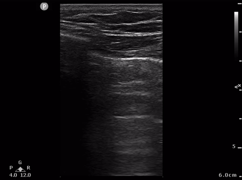
Figure 1. A-lines without lung sliding present

Figure 2.Barcode sign
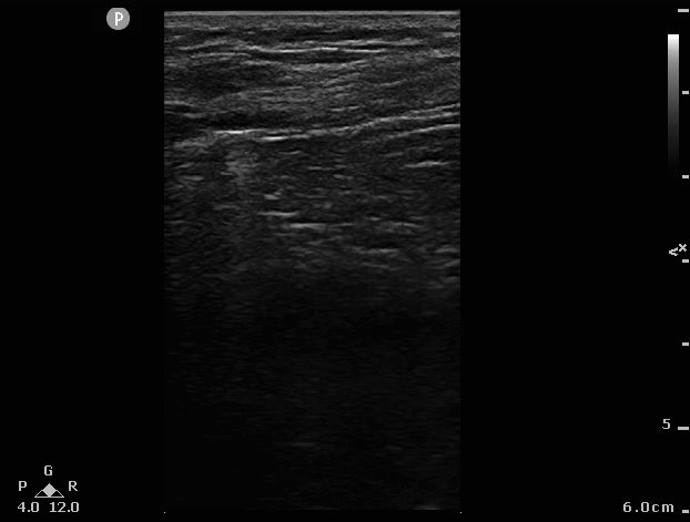
Figure 3.Lung point
References
- Vincent JL, Abraham E, Moore FA, Kochanek P, Fink MP, eds. Textbook of Critical Care. 8th edition. Elsevier; 2023.
- Marini TJ, Rubens DJ, Zhao YT, Weis J, O'Connor TP, Novak WH, Kaproth-Joslin KA. Lung Ultrasound: The Essentials. Radiol Cardiothorac Imaging. 2021 Apr 29;3(2):e200564. doi: 10.1148/ryct.2021200564. PMID: 33969313; PMCID: PMC8098095.
- Husain LF, Hagopian L, Wayman D, Baker WE, Carmody KA. Sonographic diagnosis of pneumothorax. J Emerg Trauma Shock. 2012 Jan;5(1):76-81. doi: 10.4103/0974-2700.93116. PMID: 22416161; PMCID: PMC3299161.
- Alrajab, S., Youssef, A.M., Akkus, N.I. et al. Pleural ultrasonography versus chest radiography for the diagnosis of pneumothorax: review of the literature and meta-analysis. Crit Care 17, R208 (2013). https://doi.org/10.1186/cc13016
- FAST and eFAST Ultrasound. Taming The SRU. Accessed Dec. 05, 2024. https://www.tamingthesru.com/us/scanning-school/fast
Author Affiliations
- Stuart Brill, DO; Department of Neurosurgery, Westchester Medical Center, Valhalla, New York
- Georgina Ang, MD; Department of Medicine, Westchester Medical Center, Valhalla, New York
- Jon Rosenberg; Department of Neurosurgery, Westchester Medical Center, Valhalla, New York